Photogrammetry is gaining importance in implant prosthetics — particularly in complex full-arch restorations. On four, six or more implants, the framework must be seated free of tension; without mechanical stress. The benchmark is called passive fit; and it allows no compromise.
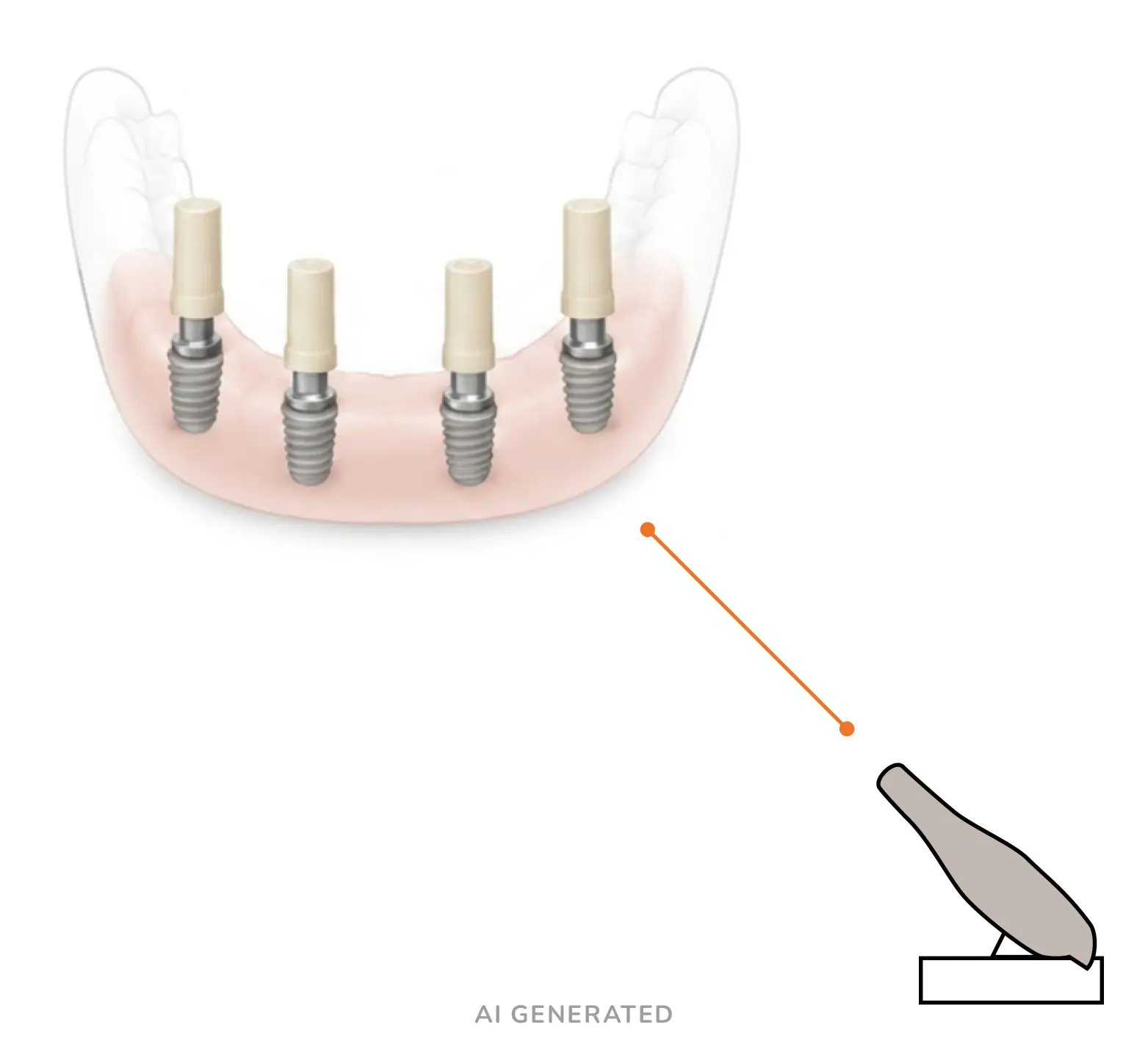
Whether passive fit is achieved in complex implant cases also depends on data acquisition and measuring technology. While intraoral scanners capture many implant restorations precisely, they reach their limits with full-arch cases. Many practices have so far solved this with conventional impression techniques. Photogrammetry offers a precise digital alternative for this.
The intraoral scanner reconstructs surfaces. Photogrammetry calculates coordinates in space. Both are needed – but for different tasks.
Why stitching becomes a challenge in full-arch cases
An intraoral scanner works with optical triangulation and stitching: the handpiece continuously captures small sections. The software assembles these into an overall image. In the dentate jaw this works very well. Distinctive geometries such as cusps, fissures and edges give the algorithm reliable anchor points.
In the edentulous jaw with several implants, the situation changes. The mucosa offers the stitching algorithm hardly any geometric reference points. If these are missing, the algorithm can drift minimally when joining the data – depending on the scanner system, scan body design and scanning strategy. Over the distance of a dental arch, the risk can add up to a measurable distortion effect: the recorded implant positions deviate from their spatial relationship to one another.
The measuring principle of photogrammetry
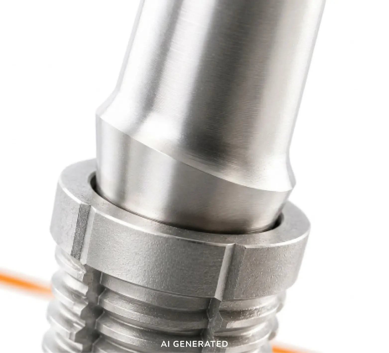
Photogrammetry is based on a different physical measuring principle. The aim is not the reconstruction of surfaces, but the determination of the spatial coordinates of implants. For this purpose, specially coded measuring bodies — the photogrammetry scan bodies — are screwed onto the implants. Their geometry is mathematically defined. A calibrated camera system captures the measuring bodies from several angles. The software brings all image points together in a joint optimisation procedure (bundle adjustment).
The algorithm calculates the position and axial inclination of each implant within the same mathematical reference system. The error accumulation typical of stitching procedures is thereby avoided.
For single implants, short spans and many partially dentate situations, modern intraoral scanners deliver clinically very good results. In full-arch restorations in particular, however, the requirements for the spatial accuracy of the implant positions increase.
Passive fit: what the figures mean
The clinically tolerable limit value for a passive fit is frequently stated in the literature as up to 150 µm. In current studies, photogrammetry systems achieve linear deviations between 10 µm and 49 µm and thus remain clearly and consistently below this critical limit value. The practical consequences:
- Freedom from tension: minimal risk of screw loosening, abutment disfit or fatigue fractures of the superstructure.
- Efficient workflow: reduced need for elaborate intermediate steps such as analogue verification jigs or milled resin prototypes for checking the fit.
- Lower dependence on implant depth: reliable geometric capture even with implants inserted deep subgingivally.
Two technologies, one workflow
Photogrammetry does not replace the conventional intraoral scanner, but complements the options. For teeth, soft tissue, the opposing jaw and bite registration, the IOS remains the right tool. For the spatial capture of several implant positions in long-span restorations, photogrammetry takes over. In the CAD software, the two data sets are merged: the coordinate skeleton of the photogrammetry is superimposed on the soft-tissue scan of the IOS. The result is an overall digital image that neither the measuring technology of the one nor that of the other procedure could provide on its own.
OrangeCAD: from the measuring body to the titanium base – precision as a constant
OrangeCAD also manufactures photogrammetry scan bodies for an established supplier of dental photogrammetry systems. The manufacture of photogrammetry scan bodies places particular demands on precision, process stability and quality assurance. At OrangeCAD, these standards are also incorporated into the production of titanium bases and pre-mill abutments.
References
Gómez-Polo M. et al. (2023): Influence of arch location and scanning pattern on the scanning accuracy, scanning time, and number of photograms of complete-arch intraoral digital implant scans. Clinical Oral Implants Research, Vol. 34(11), 1214–1226.
Fu X. et al. (2025): Intraoral Photogrammetry: The Next Step in Full-Arch Implant Precision. Dentistry Today, Vol. 44(3), 64–69.
Negreiros W. M. et al. (2025): Photogrammetry in Implant Dentistry. Australian Dental Journal, Vol. 70(Suppl 1), S15–S24.
Eldabe A. K. et al. (2025): Accuracy of intraoral photogrammetry in complete arch digital implant scanning: An in vivo prospective comparative study. The Journal of Prosthetic Dentistry, Vol. 134(4), 782–789.